- Wireless remote control of multiple units (maximum 5 units). NEW
- Playlist can be created. NEW
- Touchscreen control panel. NEW
- Ready- to-use with connecting to power supply. NEW
- Sounds are recorded from real patients and are reproduced by using a high quality sound system.
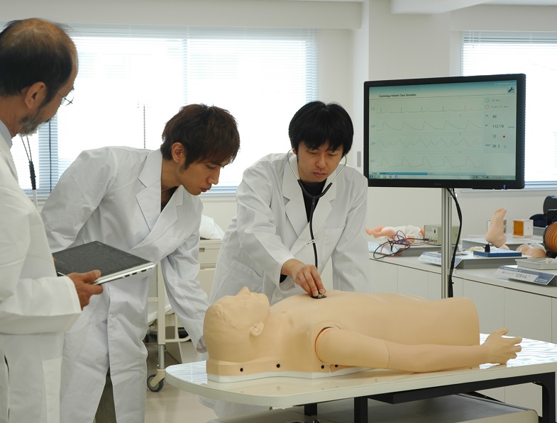
- A stethoscope can be used.
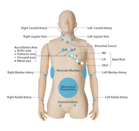
- Auscultation sites corresponding to heart valves are located on a life-size manikin body.
Real sounds, real instruments, real anatomy
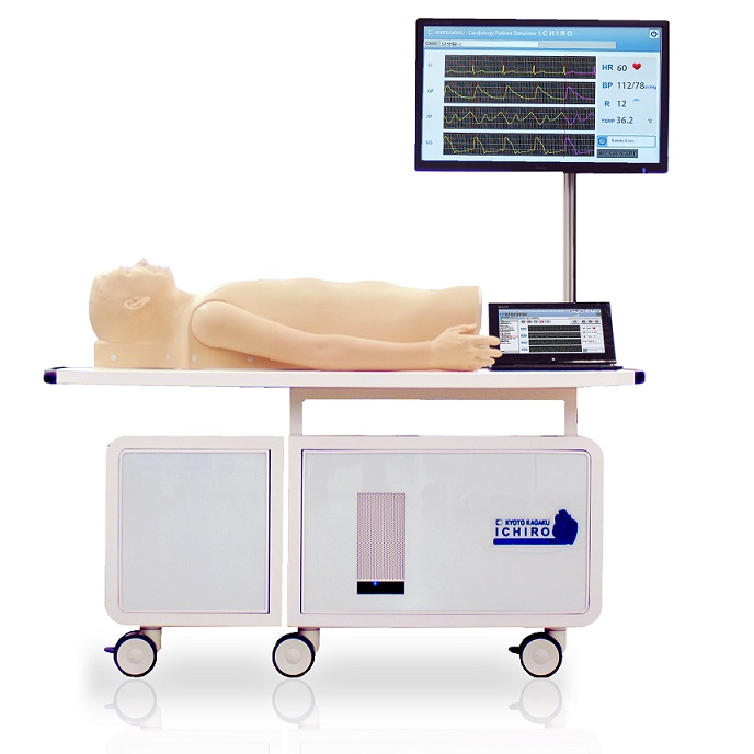
- Sounds are recorded from actual people and reproduced using a high quality sound system. An actual stethoscope can be used. Auscultation sites corresponding to heart valves are located precisely on a life-size manikin body molded from an actual person
Wide variety of the examples
- Simulator "K" contains 88 cases; 12 cases of normal heart sounds, 14 cases of heart disease simulations, 10 cases of arrhythmia simulations and 52 cases of ECG arrhythmia simulations.
Comprehensive clinical examination training
- Physical findings synchronize perfectly with each other.
Construct an original education program
- Sound volume, pulse strength, simulation speed and running time are controllable.
Observation of jugular veins
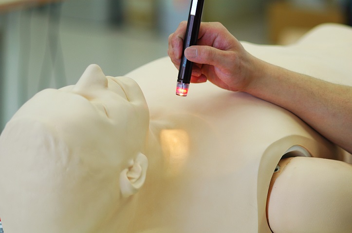
- Pulsation of jugular venous waves can be observed on both sides.
- The strength and timing of "a" and "v" waves which vary in each case can be observed just as with real cardiac patients.
Palpation of cardiac impulses (RV, LV and DLV)
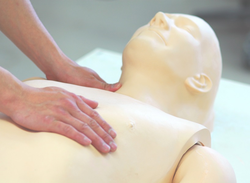
- Cardiac impulses are palpable at sites of Right Ventricle, Left Ventricle and Dilated Left Ventricle.
- Various cardiac impulses under different cardiac conditions are simulated.
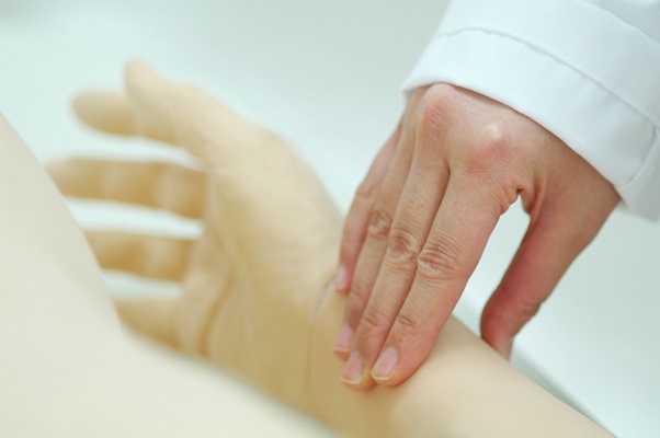
Palpation of arteries
- The carotid, medial, radial and femoral arteries are palpable at eight sites on the manikin. Slight variations of the arterial pulse waves under different cardiac conditions or arrhythmias can be detected by palpation.
Heart sounds & murmurs
- In all cases listening can be performed at the four primary cardiac auscultation sites (aortic, pulmonic, tricuspid, and mitral). Auscultation of first sound (S1) and second sound (S2) can be learned in relation to synchronized electrocardiogram, arterial pulses and jugular venous waves.
Respiratory sounds and observation of abdominal movement (for cases HR: 60/ min)
- Tracheal and bronchial breath sounds and abdominal movement are simulated to facilitate understanding of respiratory related phenomena such as Rivelo-Carvallo sign, respiratory splitting and timing of murmurs.
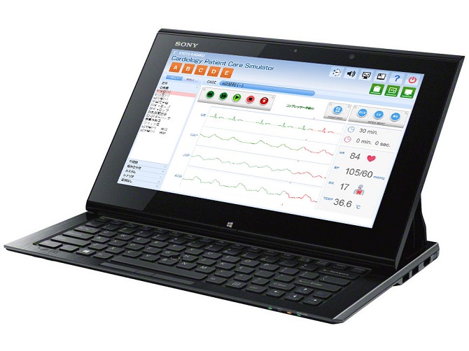
Monitoring screen
- Electrocardiogram (ECG), jugular venous pulse (JVP) and carotid arterial pulse (CAP) and apex cardiogram (ACG)
- Each chart can be freeze-framed for in-depth learning.
- Case explanation windows for self-directed learning are provided.
Group study
- External speaker system produces heart sounds respectively at each auscultation site (aortic, pulmonic, tricuspid, and mitral).
- Useful for pre-training demonstration, group discussions and problem-based learning exercises
New Features
Up to 5 Simulators at a time Playlist System OSCE, group session and scenario based simulation
Up to five cardiology simulators can be controlled by one wireless tablet.
- each simulator can be individually set with a different cardiac case
- cases can be switched promptly at any time during the session
Playlist System
- Playlist facilitates trouble-free training/OSCE session
- Choose up to 10 cardiac cases from 88 examples to make a playlist
Ready-to-use
- Simple connection and all-in-one unit structure.
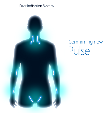
Error Indicator
- The error indicator performs maintenance of the system to keep Simulator K in its best condition.
- If there are problems that may occur in the system, an indicator will warn you on the screen.
- All system problems are recorded in the systems history.
Cases & Simulation Contents
|
Normal heart simulation (12 cases) |
Heart disease simulation (14 cases) |
Arrhythmia (10 cases) |
|---|---|---|
|
S2 split (-) HR: 60 S1 split (+) S2 split (+) S2 wide split S3 gallop S4 gallop pulmonic ejection sound S3 and S4 gallop innocent murmur midsystolic click sound S2 split (-) HR: 72 S2 split (-) HR: 84 |
aortic stenosis mitral regurgitation mitral stenosis aortic regurgitation hypertrophic cardiomyopathy mitral steno-regurgitation pulmonic valvular stenosis atrial septal defect ventricular septal defect tricuspid regurgitation acute mitral regurgitation patent ductus arteriosus mitral valvular prolapse dilated cardiomyopathy |
sinus arrhythmia sinus tachycardia sinus bradycardia ventricular premature contraction (1) ventricular premature contraction (2) ventricular premature contraction (3) sino atrial block atrio-ventricular block atrial fibrillation atrial flutter |
| Normal jugular venous waves, arterial pulses and cardiac impulses are simulated, as well as heart sounds such as S2 splitting in the pulmonic area and S3 and S4 gallop sounds in the mitral area. | The characteristic findings of the arterial and venous pulse waves are simulated. For example, in ventricular premature contraction, the venous pulsations are normal but arterial pulsation is barely palpable by the premature beat. | Characteristic heart sounds and pulse waves are simulated, Such as, ventricular premature beats. |
ECG: Arrhythmia simulation
- A-01 normal sinus R
- A-02 sinus tachycardia
- A-03 sinus arrhythmia
- A-04 apc solitary
- A-05 apc bigeminy
- A-06 ectopic pacemaker
- A-07 wondering pacemaker
- A-08 coronary sinus R
- A-09 sinus bradycardia
- A-10 s s syndrome
- A-11 atrial fibrillation
- A-12 atrial flutter
- A-13 atrial flutter fib
- B-01 atrial flutter
- B-02 av block
- B-03 av block & crbbb
- B-04 av block (digital)
- B-05 av block (mobitz)
- B-06 av block (mobitz)
- B-07 av block (3:1&4:1)
- B-08 av & crbbb
- B-09 paroxy atr tachy
- B-10 av junc R (svst)
- B-11 av junc R (pat)
- B-12 av junc R
- B-13 av junc contraction
- C-01 V VI pacemaker
- C-02 atrial pacemaker
- C-03 vent pacemaker
- C-04 av seq pacemaker
- C-05 icrbbb
- C-06 crbbb
- C-07 clbbb
- C-08 clbbb
- C-09 clbbb (by ami)
- C-10 wpw syndrome
- C-11 wpw syndrome
- C-12 wpw syndrome
- C-13 vpc (solitary)
- D-01 vpc (quadrigeminy)
- D-02 vpc (trigeminy)
- D-03 vpc (bigeminy)
- D-04 vpc (couplet)
- D-05 pvc (repetitive)
- D-06 pvc (R-on-T type)
- D-07 non-sustained VT
- D-08 vent tachycardia
- D-09 vent flutter
- D-10 vent fibrillation
- D-11 vent R (sinus cond)
- D-12 accel vent rhythm
- D-13 agonal rhythm
In addition to 36 patient simulations, the software allows in-depth study of ECG in various arrhythmias. The full size graphic ECG is displayed to practice reading the waves using pause and/or calibrator functions. Fifty-two pre-recorded cases are classified into 4 categories, comprised of 13 cases each.
Set Compnonets:
1 cardiology model unit
1 table
1 set of speaker system
1 set of PC, keyboard and mouse (these 2 sets are built into the unit and can not be removed.)
1 LCD monitor (27 inch)
1 control PC
4 textbooks
1 rib sheet
1 cover
1 instruction manual
Who is Mr. "K"?
Simulator "K" is a simulated cardiology patient for clinical training. He facilitates total training in bedside clinical examination skills and ensures quality of training in auscultation of heart sounds and murmurs. He was born in Japan in 1997.
Obtaining reliable auscultation skills
Auscultation is a fundamental approach to cardiac patients, performed widely from general practitioners to cardiologists. Repeated practice is a necessity for learners to differentiate various heart sounds and murmurs. However, opportunities to learn with real patients are limited and could be insufficient. Simulator "K" offers hands-on experience in a diversity of cases.
- Replacement plus tubes

