Home ->
Medical Education ->
Simulators -> Orthopedics -> Knee Aspiration & Injection Trainer with Ultrasound Capabilities
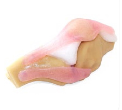
- The following key anatomical landmarks are realistic to palpate:
-
- Skin
- Subcutaneous fat, quadriceps tendon & patellar ligament
- Prefemoral fat pad
- Suprapatellar fat pad
- Hoffa (Infrapatellar) fat pad
- Femur
- Medial & lateral collateral ligament
- Tibia
- Patella
- Joint space & synovial recess
- Meniscus
- Muscle mass of quadriceps
- Discrete muscle and skin layers provide realistic tissue and needle response
- Anatomically accurate synovial sac and palpable patella
- Realistic colour and consistency of synovial fluid
- 1,000+ stabs per module with 21 gauge needle
- Precise, palpable anatomy with bony landmarks
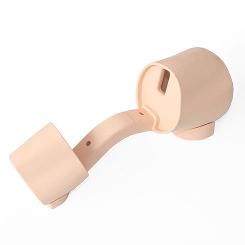
- Robust, sealed knee unit that holds all anatomy
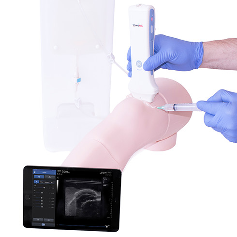
- Key internal landmarks visible under ultrasound
- Compatible with all standard ultrasound machines
- Echolucent material allows aspiration and injection under ultrasound-guidance or using the palpation method
- Suitable for Undergraduate and Postgraduate medical study
- Knee skin is watertight
- Skin washable using mild soap and water
- Can be used with leg supports for supine position, or without leg supports for side of bed position
- Caters for ultrasound-guided techniques as well as palpation
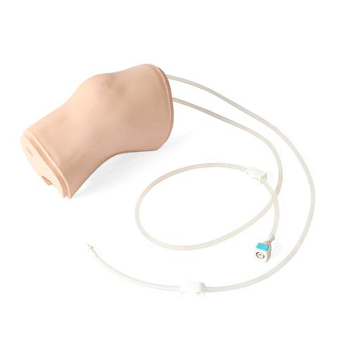
- Separate Fluid Bag & Stand ensure the model is easy to use and mobile
- Clear indicator on Fluid Bag to prevent overfilling
- Latex free
Components:
1 Ultrasound Knee Module for Aspiration & Injection
1 Fluid Bag and Stand
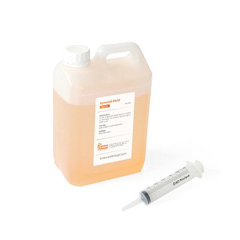
1 Synovial Fluid (including syringe)
1 Needle Set
1 Leg Unit, including Removable Supports
1 Carry Case for Knee
Joint and Soft Tissue Injection (5th Edition) By Dr Trevor Silver
- Injection into joint cavity
- Aspiration of synovial fluid from both the lateral and medial aspects
- Identification of anatomical landmarks using the palpation method or ultrasonic guidance
- Patient positioning and management
- Recognition of joint effusion
- Ballottement
- Competence using ultrasound technology to perform systematic scanning techniques and examination of the knee joint
- Synovial Fluid


