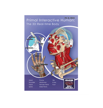
- Ultimate 3D interactive anatomy viewer and image library for all medical educators and practitioners who wish to create custom images for handouts and lectures.
- It allows the user to add their own labels and annotations and is available in English, German, Portuguese, Spanish, Latin, Japanese and Chinese.
- Accurately built from real scan data and published in an easy to use real-time interface, you can explore the human body region by region, with over 3000 structures included in the 11 models:
Head & Neck
Spine
Shoulder
Knee
Hand
Thorax
Abdomen
Foot & Ankle
Female Pelvis
Male Pelvis
Hip
- Easy to navigate to all content.
- Anatomically accurate 3D interactive models allows you to explore the human body, region by region, with over 3000 structures included in the 11 models.
- Create customised images with your own labels and annotations, then print or save them for use in your own presentations and hand outs.
- 100% user-driven functionality allows you to rotate models in any direction using your mouse, zoom in/out, and choose which anatomical structures – muscles, vessel systems and organ systems – are added or removed in groups or individually, made x-ray, opaque or viewing in isolation. What better way to learn, understand, teach and explain anatomy and the relationships between structures.
- Flexible settings to create outline-only views - great for colouring exercises - or 3D stereograph views (for a truly 3D experience!).
- Export and print content royalty-free for your own presentations, patient education and student handouts.
Technical Specification:
Requires an internet connection.
Recommended web browsers:
Internet Explorer 7 + (Windows)
Firefox 2 (on Windows)
Safari (Mac)
Chrome
* Your login and password will be sent you by email on completion of your order.


